
STRUCTURAL ANALYSIS OF MATERIALS
- CRYSTALLOGRAPHIC ANALYSIS
- RADIOGRAPHIC ANALYSIS
- ELECTRON MICROSCOPY
- OPTICAL MICROSCOPY
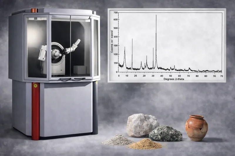
Crystallographic analysis of materials is performed in the Laboratory using Powder X-ray Diffraction (XRD). The technique is based on the interaction of X-rays with the crystal structure of materials. When an X-ray beam strikes a crystalline material, atoms arranged in periodic crystal planes diffract the radiation at specific angles. The resulting diffraction pattern constitutes a characteristic fingerprint of the material’s crystal structure.
The measurement follows Bragg’s law, according to which diffraction angles are directly related to the distances between crystal planes. Analysis of the intensity-angle diagram (diffractogram) enables the crystalline phases present in a sample to be identified.
Powder XRD requires a small quantity of material to be collected and ground into a powder before measurement.
Information Provided by the Analysis
Crystallographic analysis can determine:
- the mineralogical or crystalline composition of a material
- the identity of crystalline phases
- the presence of multiple phases in composite materials
- the degree of crystallinity
- possible structural changes or phase transformations
- average crystallite size and microstructural parameters in specialised analyses
Materials That Can Be Analysed
The method can be applied to a wide range of materials, including:
- minerals and geological samples
- ceramics and building materials
- archaeological materials (ceramics, mortars, pigments)
- metallic and industrial materials
- environmental samples (sediments, soils, dusts)
- advanced and composite materials
Applications
Crystallographic analysis is used in numerous research and applied studies, including:
- archaeometric studies and characterisation of archaeological materials
- mineralogical analysis of geological and environmental samples
- investigation of ceramics and mortars
- qualitative and quantitative phase analysis of industrial materials
- investigation of phase transformations caused by thermal or chemical processes
- material characterisation in materials and nanomaterials research
Radiographic analysis of materials is performed in the Laboratory using digital X-ray radiography. This non-destructive testing technique enables the internal structure of an object to be imaged without sampling or intervention in the material.
The method is based on the differential absorption of X-rays by materials. When an X-ray beam passes through an object, part of the radiation is absorbed and part is transmitted through the material. The recorded radiation intensity depends on the thickness, density, and composition of the materials. In digital radiography, the radiation emerging from the object is recorded by a digital detector, which converts the signal into a high-resolution digital image.
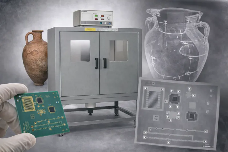
Digital technology enables immediate image display, processing, and analysis, as well as enhancement of contrast and sharpness to reveal details of the internal structure.
X-ray radiography is a non-destructive technique because the object is examined without sampling or any physical alteration.
Information Provided by the Analysis
Radiographic examination can reveal:
- the internal structure of an object
- cracks, voids, and internal discontinuities
- the presence of different materials or layers
- manufacturing and assembly techniques
- inclusions or foreign bodies within a material
- evidence of previous interventions or repairs
Materials That Can Be Analysed
The technique can be applied to a wide range of materials, including:
- archaeological objects (ceramics, metal objects, coins, tools)
- works of art and cultural heritage objects
- geological and mineralogical samples
- ceramics and building materials
- metallic and composite materials
- industrial products
Applications
Radiographic analysis is used in numerous research and applied studies, including:
- archaeometric and museum studies aimed at understanding object-manufacturing technology
- authenticity assessment and detection of subsequent interventions
- detection of internal deterioration or structural alteration in cultural heritage objects, such as paintings, wooden objects, sculptures, and other works of art
- non-destructive testing of materials in industrial applications
- investigation of structural discontinuities or defects
- investigation of the internal structure of composite objects
Electron microscopy is an advanced structural-analysis technique that enables the morphology and microstructure of a sample to be observed at very high magnification. The Laboratory uses a variable-pressure Scanning Electron Microscope (SEM) equipped with multiple detectors and advanced analytical systems.
SEM operation is based on scanning the sample surface with a finely focused electron beam. The interaction of the electrons with the material produces various signals, including secondary electrons, backscattered electrons, and characteristic X-rays. These signals are recorded by specialised detectors and converted into high-resolution images and analytical data.
Electron microscopy is generally regarded as a non-destructive technique, as only a small quantity of sample is required for analysis. In some cases, limited sample preparation may be necessary, without substantially altering its structure.
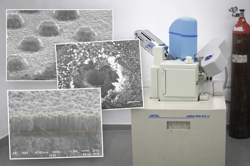
A major advantage of the APML’s SEM is its ability to operate under variable-vacuum conditions (low or high vacuum). This enables non-conductive materials to be examined without necessarily coating them with a conductive material, such as gold or carbon, as is required in conventional high-vacuum SEM. Natural samples, including ceramics, soils, minerals, and biological materials, can therefore be observed directly without extensive preparation.
Capabilities and Information Provided
Electron microscopy can be used to investigate:
- surface morphology and microstructure
- the distribution of grains and crystals
- microcracks, pores, and other structural discontinuities
- surface microtopography and texture
- the presence of inclusions or secondary phases
Materials That Can Be Analysed
The technique can be applied to a wide range of materials, including:
- archaeological and archaeometric materials (ceramics, metals, glass, mortars)
- geological and mineralogical samples
- metallic materials and alloys
- ceramics and composite materials
- industrial materials and advanced technological materials
- environmental samples (soils, sediments, particles)
Advanced Analytical Capabilities
-
Secondary-electron (SE) detector
for detailed imaging of surface morphology -
Backscattered-electron (BSE) detector
for highlighting differences in chemical composition and phase density -
EDS (Energy-Dispersive X-ray Spectroscopy)
for qualitative and quantitative elemental microanalysis -
WDS (Wavelength-Dispersive X-ray Spectroscopy)
for higher-accuracy elemental determination -
EBSD (Electron Backscatter Diffraction)
for investigating crystal structure and crystallographic orientation
Applications
Electron microscopy is widely used for:
- archaeometric studies investigating the manufacturing technologies of archaeological materials
- investigation of the microstructure of ceramics, metals, and glass
- characterisation of minerals and geological materials
- investigation of corrosion and material deterioration
- analysis of particles and microstructures in environmental samples
- research and development of new materials and industrial applications
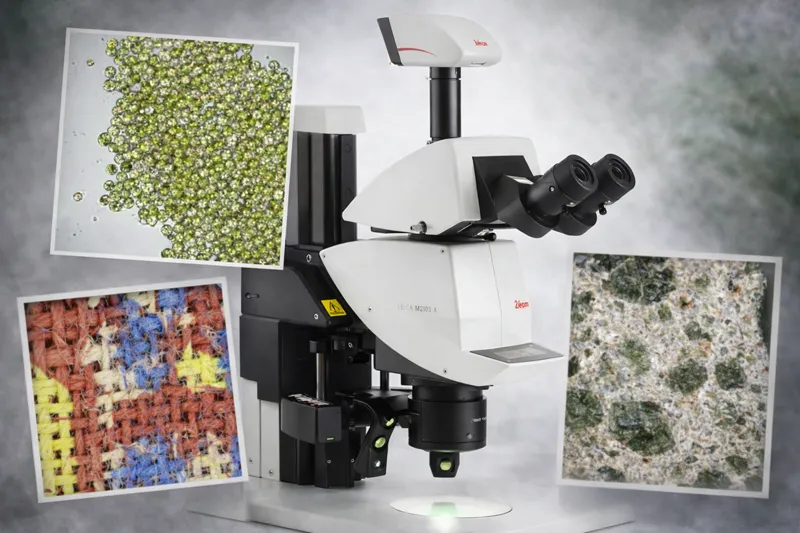
Optical microscopy is a fundamental technique for observing and analysing material structure at the microscale. The Laboratory uses modern optical-microscopy systems, including an automated stereomicroscope and a high-resolution biological microscope with phase-contrast capability, both equipped with digital cameras for image acquisition and documentation of observations.
The technique is based on observing the interaction of visible light with the sample, enabling the morphology, microstructure, and surface characteristics of materials to be imaged. Different magnifications and illumination techniques allow detailed examination of the surface or the internal structure of thin samples.
Optical microscopy is a non-destructive technique, as it usually requires little or no special sample preparation and enables direct observation without altering the material’s structure. In some cases, simple preparation, such as sectioning or surface polishing, may be required.
Capabilities and Information Provided
Optical microscopy can reveal:
- the morphology and surface microstructure of materials
- texture and microtopography
- microcracks, pores, and surface deterioration
- the distribution of grains, crystals, or particles
- inclusions and heterogeneities within a material
- biological structures, such as cells, microorganisms, and other microscopic organisms
Materials That Can Be Analysed
Optical microscopy can be applied to a wide range of materials, including:
- archaeological and archaeometric materials (ceramics, mortars, pigments, fibres)
- geological and mineralogical samples
- metallic and ceramic materials
- biological materials (cells, microorganisms, plant tissues)
- environmental samples (soils, sediments, particles)
- food and agricultural products
Advantages of the Laboratory Systems
-
The automated stereomicroscope
enables three-dimensional observation of object surfaces at low and medium magnifications, providing excellent perception of sample morphology and topography. This technique is particularly useful for examining objects without special preparation. -
The phase-contrast microscope
enables transparent or semi-transparent samples to be observed, revealing fine structures that are not readily visible under conventional illumination. Phase-contrast microscopy converts small differences in the phase of light passing through the sample into intensity differences, improving the visualisation of fine microstructures.
Applications
Optical microscopy is widely used for:
- archaeometric studies investigating material structure and manufacturing technology
- identification of microstructures and textures in ceramic or geological materials
- investigation of deterioration and surface alteration
- observation of particles and fibers in environmental samples
- microscopic observation of biological samples and microorganisms
- preliminary microscopic examination before more specialised analytical techniques, such as SEM
Optical microscopy is particularly useful for the initial microstructural investigation of materials and biological samples, providing a rapid and reliable overview of their morphology and characteristics.